Spring Sale Special Limited Time 70% Discount Offer - Ends in 0d 00h 00m 00s - Coupon code: buysanta

Exact2Pass Menu

Musculoskeletal Ultrasound Certification in Rheumatology

Last Update 19 hours ago Total Questions : 100

The Musculoskeletal Ultrasound Certification in Rheumatology content is now fully updated, with all current exam questions added 19 hours ago. Deciding to include RhMSUS practice exam questions in your study plan goes far beyond basic test preparation.

You'll find that our RhMSUS exam questions frequently feature detailed scenarios and practical problem-solving exercises that directly mirror industry challenges. Engaging with these RhMSUS sample sets allows you to effectively manage your time and pace yourself, giving you the ability to finish any Musculoskeletal Ultrasound Certification in Rheumatology practice test comfortably within the allotted time.

Question # 11

Which probe is the BEST choice for ultrasound examination of the adult wrist?

A.

Linear 20-25 mm, 3-8 MHz transducer

B.

Linear 20-25 mm, 10-18 MHz transducer

C.

Linear 40-50 mm, 3-8 MHz transducer

D.

Linear 40-50 mm, 10-18 MHz transducer

Question # 12

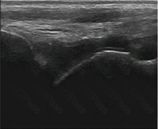
Which pathologic condition is MOST likely responsible for the etiology of shoulder pain in the patient?

A.

Rheumatoid synovitis

B.

Osteoarthritis

C.

Calcific tendinitis

D.

Subacromial bursitis

Question # 13

Which of the following protocols would be MOST consistent with a complete routine ultrasound examination of the shoulder?

A.

Anterior long- and short-axis views of the biceps tendon

o Anterior view of the subscapularis tendon in external rotation

o Long- and short-axis views of the supraspinatus tendon, moving the transducer anteriorly and posteriorly

o Posterolateral view of the infraspinatus tendon

o Posterior view of the glenohumeral joint recess

o View from cranial at the acromioclavicular joint

B.

Anterior long- and short-axis views of the biceps tendon

o Anterior view of the rotator cuff interval

o Lateral views of the subscapularis tendon

o Lateral long- and short-axis views of the supraspinatus tendon, moving the transducer anteriorly and posteriorly

o Posterior view of the glenohumeral joint recess

o Dynamic lateral long-axis assessment of impingement with abduction of the arm

C.

Anterior long- and short-axis views of the biceps tendon

o Anterior views of the pectoralis tendon insertion

o Anterior view of the coracoid ligament and rotator cuff interval

o Long- and short-axis views of the supraspinatus tendon, moving the transducer anteriorly and posteriorly

o Posterior long- and short-axis views of the infraspinatus tendon insertion

o Axillary long-axis view with arm raised

D.

Anterior long- and short-axis views of the biceps tendon

o Anterior view of the rotator cuff interval

o Long- and short-axis views of the supraspinatus tendon, moving the transducer anteriorly and posteriorly

o Dynamic lateral long-axis assessment of impingement with abduction of the arm

o Posterior transverse view of the suprascapular notch

o View from cranial at the acromioclavicular joint

Question # 14

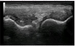
This ultrasound image from a 60-year-old woman with knee pain is MOST suggestive of which of the following?

A.

Urate deposition

B.

Meniscus tear

C.

Chondrocalcinosis

D.

Osteophyte formation

Question # 15

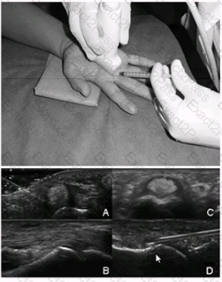
A photograph of an ultrasound-guided injection approach is shown below. With which ultrasound image does it BEST correlate?

A.

Image A

B.

Image B

C.

Image C

D.

Image D

Question # 16

Ultrasound evaluation of a painful shoulder is MOST likely to be useful in planning a corticosteroid injection for which of the following conditions?

A.

Complex regional pain syndrome

B.

Adhesive capsulitis

C.

Superior labral anterior-posterior lesion

D.

Glenohumeral synovitis

Question # 17

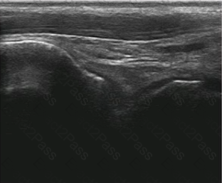
A practitioner wants to assess the structures on the right, distal portion of this image.

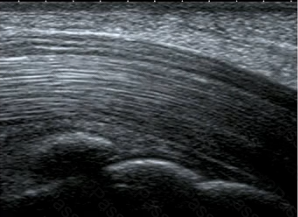
What should the practitioner do at this time?

A.

Choose a lower transducer frequency.

B.

Place the focal point at the level of the area of interest.

C.

Increase the overall gain to brighten the image.

D.

Depress the distal end of the probe.

Question # 18

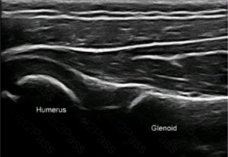
This posterior transverse scan of the shoulder of a 3-year-old boy shows which of the following?

A.

Effusion of the glenohumeral joint

B.

Effusion with an erosion of the humeral head

C.

Normal shoulder scan

D.

Fracture of the humeral head

Question # 19

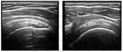
The change between the ankle depicted in the first and second images would BEST be explained by which of the following?

A.

Decreased synovitis

B.

Decreased transducer pressure

C.

Decreased frequency

D.

Increased gain

Question # 20

At which angle is a dorsal scan of the elbow BEST performed?

A.

Flexion of the elbow joint in a 45-degree angle

B.

Flexion of the elbow joint in a 60-degree angle

C.

Flexion of the elbow joint in a 90-degree angle

D.

Full extension of the elbow joint

Go to page: