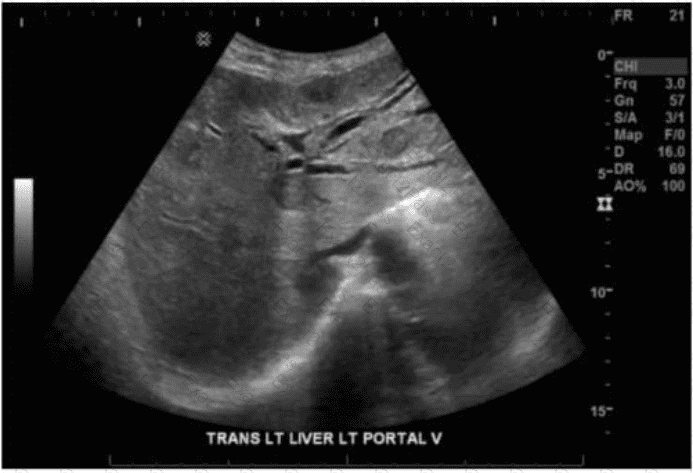
Which sonographic appearance of the bile ducts is demonstrated in this image?
Normal intrahepatic
Dilated intrahepatic
Dilated common bile
Dilated common hepatic
The image shows a transverse view of the left lobe of the liver with the portal triads clearly visible. The "parallel channel" or "double barrel" sign is observed—where dilated intrahepatic bile ducts run alongside the portal veins, creating a characteristic sonographic pattern of paired anechoic (black) tubular structures.
This sonographic feature is diagnostic for dilated intrahepatic bile ducts and is typically seen in obstructive jaundice or biliary obstruction from conditions such as:
Choledocholithiasis (stone in the common bile duct)
Stricture or mass compressing the bile ducts
Cholangiocarcinoma
The intrahepatic bile ducts normally are too small to visualize clearly unless dilated. Their dilation gives the liver a "too many tubes" appearance, where bile ducts become as prominent as the portal veins.
Comparison of answer choices:
A. Normal intrahepatic ducts are not usually seen this clearly or prominently on ultrasound.
B. Dilated intrahepatic — Correct. The parallel channel sign supports this diagnosis.
C. Dilated common bile duct would be visualized extrahepatically, typically anterior to the portal vein near the head of the pancreas.
D. Dilated common hepatic duct is also extrahepatic and seen in the porta hepatis, not within the liver parenchyma.
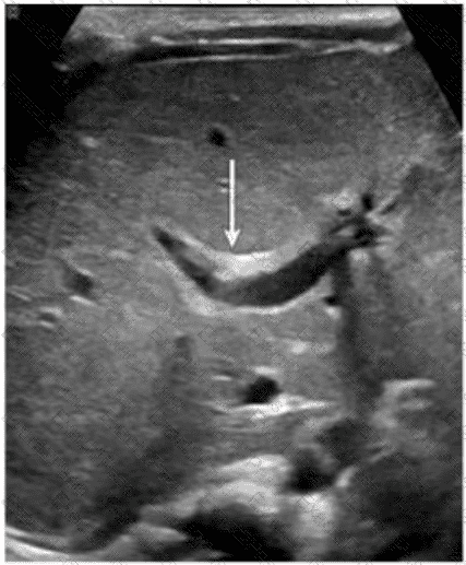
Which congenital disorder is most consistent with the finding identified by the arrow on this image?
Sclerosing cholangitis
Alagille syndrome
Caroli disease
Biliary atresia
The image demonstrates a characteristic "central dot sign" — a hallmark finding of Caroli disease. This is best appreciated on ultrasound as a cystic dilation of the intrahepatic bile ducts with a central echogenic dot or linear structure (which corresponds to the portal vein and fibrous tissue within the dilated duct). The arrow in the image points to one such dilated duct.
Caroli disease is a rare congenital disorder characterized by segmental, saccular dilation of intrahepatic bile ducts. It is often associated with congenital hepatic fibrosis and may predispose to cholangitis, stone formation, and even cholangiocarcinoma.
Key ultrasound features of Caroli disease:
Cystic or saccular dilations of the intrahepatic bile ducts
The "central dot sign" — echogenic focus in the center of the dilated ducts (representing portal vein radicle or fibrous tissue)
May show associated hepatosplenomegaly or signs of portal hypertension
Differentiation from other options:
A. Sclerosing cholangitis: Typically causes diffuse or segmental biliary ductal wall thickening and stricturing; does not present with cystic dilations.
B. Alagille syndrome: A multisystem disorder often characterized by a paucity of intrahepatic bile ducts, not dilation.
D. Biliary atresia: Presents in infancy with obliteration of extrahepatic bile ducts, echogenic "triangular cord" sign, and absence of a visible gallbladder. It does not cause ductal dilation.
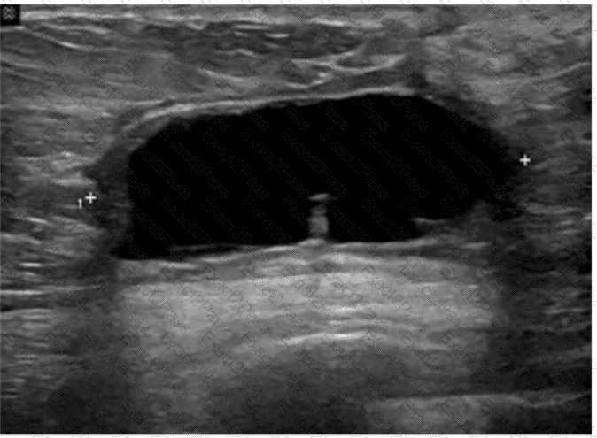
Which condition is most consistent with this image of a postsurgical breast?
Carcinoma
Blood clot
Abscess
Seroma
The ultrasound image reveals a well-defined, anechoic (black), thin-walled fluid collection located in the subcutaneous or parenchymal plane of the breast. This is most consistent with a seroma, particularly in the context of recent breast surgery.
A seroma is a common postsurgical finding, representing a sterile collection of serous fluid that accumulates in the surgical bed. It typically appears:
Anechoic (or hypoechoic if older)
Well circumscribed
Without internal septations or debris
Lacking hyperemia or surrounding inflammatory changes
This contrasts with:
A. Carcinoma — typically presents as an irregular, hypoechoic mass with angular margins, internal vascularity, and shadowing.
B. Blood clot (hematoma) — often appears heterogeneous, with internal echoes and variable echotexture depending on the age of the clot.
C. Abscess — appears as a complex fluid collection with thick walls, internal debris, septations, and surrounding hyperemia (often with clinical signs of infection).
D. Seroma — Correct. The described anechoic, clean-walled fluid collection is classic for a postoperative seroma.
Which of the following must be sterile for a percutaneous procedure?
Transducer
Machine controls
Transducer cover
Gel within transducer cover
In percutaneous procedures such as biopsies or drainages, maintaining a sterile field is critical to avoid introducing infection. While the transducer is covered by a sterile cover, the gel placed inside this cover (between the probe and the cover) must also be sterile, as it contacts the sterile field. The transducer itself and machine controls are not sterile but are handled appropriately to avoid field contamination.
According to the AIUM Guidelines:
"Sterile coupling gel must be used inside the sterile probe cover during all invasive or percutaneous procedures." (AIUM Guidelines for Cleaning and Preparing Ultrasound Transducers, 2021).
Which retroperitoneal finding is most likely associated with trauma?
Neuroblastoma
Fibrosis
Urinoma
Adenoma
Urinomas are collections of urine in the retroperitoneum that result from trauma, surgery, or obstruction causing urine leakage. Trauma is a frequent cause of urinoma formation due to disruption of the urinary tract.
According to Rumack’s Diagnostic Ultrasound:
“Urinomas may develop as a complication of trauma, surgery, or obstructive uropathy with urinary extravasation into the retroperitoneum.”
Which arteries are the immediate branches of the celiac trunk?
Proper hepatic, splenic, and supraduodenal
Common hepatic, splenic, and left gastric
Common hepatic, splenic, and right gastric
Proper hepatic, splenic, and gastroduodenal
The celiac trunk arises from the abdominal aorta and immediately divides into three primary branches:
Left gastric artery
Common hepatic artery
Splenic artery
The proper hepatic and gastroduodenal arteries are secondary branches of the common hepatic artery.
According to Moore’s Clinically Oriented Anatomy:
“The celiac trunk trifurcates into the left gastric, common hepatic, and splenic arteries.”
Which vessel is located directly proximal to the origination of the renal arteries?
Left portal vein
Splenic vein
Hepatic artery
Superior mesenteric artery
The renal arteries originate from the abdominal aorta just inferior to the superior mesenteric artery (SMA). The SMA arises anteriorly from the abdominal aorta at the level of L1, and just below it, the renal arteries branch laterally. The splenic vein, portal vein, and hepatic artery are located more superiorly in relation to the renal arteries.
According to Moore's Clinically Oriented Anatomy:
"The superior mesenteric artery arises from the anterior surface of the abdominal aorta just above the renal arteries." (Moore KL et al., Clinically Oriented Anatomy, 8th ed.)
Which renal anomaly is demonstrated on this image?
Duplicated collecting system
Crossed renal ectopia
Horseshoe kidney
Pelvic kidney
The ultrasound image labeled “SAG RUQ KIDNEY” demonstrates a midline sagittal view showing a renal parenchymal structure that extends across the midline anterior to the aorta and vertebral bodies, suggesting the presence of a horseshoe kidney.
A horseshoe kidney is a congenital renal anomaly in which the lower poles of both kidneys are fused across the midline by a parenchymal or fibrous isthmus. This isthmus typically lies anterior to the aorta and inferior vena cava and can be seen as a hypoechoic band of tissue crossing the midline on ultrasound.
Ultrasound findings characteristic of a horseshoe kidney:
Abnormally low position of the kidneys in the abdomen
Renal tissue (isthmus) bridging the lower poles anterior to the great vessels
Renal axes may be more horizontal than usual
Kidneys may appear closer together or “kissing” the spine anteriorly
Differentiation from other options:
A. Duplicated collecting system: Manifests as two separate collecting systems within one kidney, often with a central renal sinus split into two — not typically midline bridging.
B. Crossed renal ectopia: Involves one kidney crossing midline and fusing with the other on the opposite side, but they do not form a midline isthmus.
D. Pelvic kidney: A single kidney located in the pelvis due to failed ascent — it does not appear as midline fusion of two kidneys.
Which outcome would be present if the sample volume gate is larger than the examined vessel?
Indeterminate flow direction
Spike turbulence
Spectral noise
Aliasing
When the sample volume (gate) is too large, it captures signals from both the vessel and surrounding tissues or adjacent flows. This leads to a broadening of the spectral waveform and produces "spectral noise" or "spectral broadening," reducing the accuracy of velocity measurements and waveform analysis. Aliasing results from high velocity relative to the Nyquist limit, not from gate size.
According to Zwiebel’s Introduction to Vascular Ultrasound:
“Increasing the sample volume beyond the vessel size causes spectral broadening, resulting in spectral noise and inaccurate Doppler measurements.”
Which finding is an indication for renal biopsy to assess for renal failure?
Proteinuria
Leukocytosis
Hypercalcemia
Hematuria
Significant proteinuria, especially if persistent or in the nephrotic range, may indicate glomerular disease and is a common indication for renal biopsy. Leukocytosis and hypercalcemia are not specific for renal biopsy. Hematuria may warrant biopsy if accompanied by proteinuria.
According to KDIGO Clinical Practice Guidelines:
“Persistent proteinuria is one of the strongest indications for renal biopsy to evaluate underlying glomerular pathology.”
Which of the following is the most common symptom of cholelithiasis?
Jaundice
Flank pain
Biliary colic
Hemoptysis
The most common symptom of cholelithiasis (gallstones) is biliary colic, characterized by intermittent right upper quadrant or epigastric pain often triggered by fatty meals. Jaundice occurs in complications such as choledocholithiasis.
According to Rumack’s Diagnostic Ultrasound:
“Gallstones may cause biliary colic — episodic upper abdominal pain often related to food intake.”
Which probe frequency is most appropriate for imaging of the salivary glands?
2 MHz
4 MHz
8 MHz
12 MHz
Salivary glands are superficial structures, and high-frequency transducers (10–15 MHz) are optimal to obtain high spatial resolution. Lower frequencies are inappropriate as they lack sufficient resolution for superficial structures. A 12 MHz transducer provides excellent detail necessary for detecting small lesions, duct abnormalities, and vascular structures.
According to Rumack et al., Diagnostic Ultrasound:
"High-frequency linear transducers (10–15 MHz) are recommended for evaluating superficial structures such as salivary glands." (Rumack CM et al., Diagnostic Ultrasound, 5th ed.).
Which vessel is indicated by the arrow on this image?
Proper hepatic artery
Superior mesenteric artery
Left renal vein
Right renal artery
The ultrasound image demonstrates a transverse view of the abdominal vasculature, where the arrow is pointing to a circular vascular structure anterior to the aorta and posterior to the body of the pancreas — consistent with the superior mesenteric artery (SMA).
The SMA originates from the anterior aspect of the abdominal aorta just below the level of the celiac trunk and courses anterior to the left renal vein and uncinate process of the pancreas. On transverse ultrasound, it is often seen in cross-section as a round, pulsatile structure with echogenic walls, situated just anterior to the aorta. This appearance is known as the “target sign” or “bull's-eye” appearance.
Vessel Position Landmarks (transverse plane):
Aorta: Posterior and central
SMA: Just anterior to the aorta
Left renal vein: Passes between the aorta and SMA (nutcracker location)
Right renal artery: Courses posterior to the IVC toward the right kidney
Differentiation from other options:
A. Proper hepatic artery: Typically visualized within the liver hilum (portal triad), not in this anatomic location.
C. Left renal vein: Seen in transverse as a longer, oval structure crossing anterior to the aorta and posterior to the SMA.
D. Right renal artery: Arises laterally from the aorta and courses posterior to the IVC — not visualized in this axial midline location.
Which technique may provide better visualization of the common bile duct in a patient with hepatic steatosis?
Decrease overall gain
Increase dynamic range
Scan patient after a fatty meal
Decrease transducer frequency
In hepatic steatosis (fatty liver), increased echogenicity can obscure visualization of deeper structures like the common bile duct. Lowering the transducer frequency increases sound wave penetration, allowing better visualization of deep structures despite increased liver echogenicity. Decreasing gain or increasing dynamic range primarily adjusts image brightness and contrast but does not improve penetration.
According to Rumack’s Diagnostic Ultrasound:
“Lower frequency transducers are used to improve penetration and visualization of deeper structures in patients with fatty liver.”
Which diagnosis is most accurate based on the findings in this image from an adult patient?
Nephroblastoma
Clear cell carcinoma
Renal cell carcinoma
Transitional cell carcinoma
The ultrasound images (sagittal and transverse views of the left kidney) demonstrate a large, well-defined, heterogeneous mass within the renal parenchyma. This is highly characteristic of renal cell carcinoma (RCC), the most common primary renal malignancy in adults.
Renal cell carcinoma accounts for approximately 85% of all malignant renal tumors in adults. RCC often appears as:
A solid, heterogeneous, hypoechoic to isoechoic mass within the kidney
May contain areas of necrosis or hemorrhage (seen as mixed echogenicity)
Distortion of the normal renal contour
May have internal vascularity on Doppler imaging
Clear cell carcinoma (choice B) is the most common histological subtype of RCC but is not a separate diagnosis from RCC in imaging terms. Therefore, the most accurate answer is choice C: Renal cell carcinoma.
Differentiation from other options:
A. Nephroblastoma (Wilms tumor): A pediatric renal tumor, typically seen in children under 5 years of age—not applicable in adults.
B. Clear cell carcinoma: Histological subtype of RCC, not a distinct radiologic diagnosis.
D. Transitional cell carcinoma: Arises from the renal pelvis or ureter, typically appears as a central or collecting system mass rather than a cortical/parenchymal one.
Which technique is used to demonstrate the finding in this video?
Compression
Valsalva
Exhalation
Deep inspiration
The technique shown in the video is compression. In ultrasound imaging—especially of soft tissue masses, the bowel, or venous structures—compression is used to evaluate the compressibility of structures. The image demonstrates a classic grayscale ultrasound view of a lesion or structure being compressed with the probe.
Compression sonography is particularly important in:
Evaluating venous patency (e.g., for deep vein thrombosis)
Differentiating cystic from solid structures
Evaluating bowel wall abnormalities or intussusception
Assessing lymph nodes and soft tissue masses (as shown here)
When a structure compresses easily under probe pressure, it suggests that the lesion is fluid-filled or soft. In contrast, incompressibility may indicate a solid mass or thrombus.
Differentiation from other options:
B. Valsalva: Involves forced expiration against a closed airway, used primarily to assess venous reflux or inguinal hernias—not what is demonstrated here.
C. Exhalation: A respiratory maneuver that passively alters thoracoabdominal pressure, not actively performed by the operator or causing focal structural change.
D. Deep inspiration: Used to improve visualization of the liver, diaphragm, or gallbladder—not to evaluate the compressibility of soft tissue.
Which abnormality is depicted in this image of a patient who presents with a fever following a liver biopsy?
Cyst
Biloma
Abscess
Hematoma
The sonographic image shows a complex fluid collection within the liver parenchyma, with internal echoes and possibly septations, consistent with an abscess. In the clinical context of post-procedural fever following a liver biopsy, a liver abscess is the most likely diagnosis.
A liver abscess appears on ultrasound as a hypoechoic or complex fluid collection that may contain internal debris, septations, or gas (which may produce reverberation artifacts). These features distinguish it from other post-procedural complications.
A cyst (Option A) typically appears as an anechoic, well-defined lesion with posterior acoustic enhancement and no internal debris—this does not match the image or clinical setting.
A biloma (Option B) is a bile collection that can appear similar to a cyst or fluid collection but typically occurs due to bile leak; however, fever and internal complexity on ultrasound more strongly suggest abscess.
A hematoma (Option D) may also appear complex but usually presents with pain and not fever unless secondarily infected. Over time, hematomas evolve in appearance but lack septations and gas unless superinfected.
Which finding is demonstrated in this image?
Acute hepatitis
Hepatic steatosis
Medullary sponge kidney
Acute medical renal disease
The ultrasound image demonstrates diffuse increased echogenicity of the liver parenchyma with posterior beam attenuation (acoustic shadowing), findings that are consistent with hepatic steatosis (fatty liver disease). The liver appears brighter than normal, and the vascular markings, particularly of the portal veins, are obscured due to the increased parenchymal echogenicity.
Hepatic steatosis refers to the abnormal accumulation of fat within hepatocytes and is commonly associated with obesity, diabetes, alcohol use, and metabolic syndrome.
Classic sonographic features of hepatic steatosis include:
Diffuse hyperechogenicity (“bright liver”)
Poor visualization of intrahepatic vessels and diaphragm
Posterior acoustic attenuation
Increased hepatic echogenicity relative to the renal cortex
Differentiation from other options:
A. Acute hepatitis: Usually presents with normal or slightly decreased echogenicity, "starry sky" appearance due to prominent portal triads and periportal edema.
C. Medullary sponge kidney: A renal condition with echogenic medullary pyramids, not hepatic.
D. Acute medical renal disease: Affects the kidneys, often with bilateral renal enlargement and increased cortical echogenicity—again not related to liver imaging.
Which condition is most likely associated with this image of the common bile duct?
Liver mass
Cystic duct stone
Pancreatic head mass
Gallbladder stones
The ultrasound image demonstrates a dilated common bile duct (CBD), measuring approximately 7.7 mm in diameter. A normal CBD should generally measure less than 6 mm in a patient under 60 years old and may increase approximately 1 mm per decade thereafter or after cholecystectomy.
In the absence of gallstones within the CBD, one of the most concerning causes of CBD dilation is distal obstruction due to an extrinsic compressive lesion. The most common and clinically significant cause of distal CBD obstruction is a mass at the head of the pancreas.
A pancreatic head mass (e.g., adenocarcinoma) may compress the distal CBD and pancreatic duct simultaneously, resulting in the “double duct sign” — dilation of both the CBD and pancreatic duct. This is a classic finding in pancreatic cancer.
Comparison of answer choices:
A. Liver mass — unlikely to cause isolated CBD dilation unless invading the porta hepatis.
B. Cystic duct stone — may cause gallbladder hydrops but typically not CBD dilation unless Mirizzi syndrome is present.
C. Pancreatic head mass — Correct. This is the most likely cause of painless progressive CBD dilation without visible intraductal stones.
D. Gallbladder stones — These may be associated with biliary colic or cholecystitis but typically do not cause CBD dilation unless the stone has migrated and obstructed the distal duct.
Which condition is a common cause of biliary duct obstruction?
Tumor
Cholecystitis
Pneumobilia
Hepatitis
A tumor (such as cholangiocarcinoma, pancreatic head carcinoma, or metastases) is a common cause of biliary duct obstruction. It can compress or invade the bile ducts, leading to intrahepatic and extrahepatic duct dilatation.
Cholecystitis (B) typically affects the gallbladder but may rarely cause duct obstruction if complicated.
Pneumobilia (C) refers to air in the biliary tree, not obstruction.
Hepatitis (D) causes liver inflammation but not mechanical biliary obstruction.
Reference Extracts:
Rumack CM, Wilson SR, Charboneau JW, Levine D. Diagnostic Ultrasound. 5th ed. Elsevier, 2017.
Gore RM, Levine MS. Textbook of Gastrointestinal Radiology. 4th ed. Saunders, 2015.
—
Which complication would be associated with retroperitoneal fibrosis?
Aortic stenosis
Portal hypertension
Venous thrombosis
Hydronephrosis
Retroperitoneal fibrosis can encase and compress the ureters, leading to obstructive uropathy and hydronephrosis. It may also involve other retroperitoneal structures but hydronephrosis is the most common significant complication.
According to Rumack’s Diagnostic Ultrasound:
“Retroperitoneal fibrosis frequently results in ureteral obstruction, leading to hydronephrosis.”
Which vascular condition is most consistent with patent cutaneous para-umbilical channels and portal hypertension?
Caput medusae
Esophageal varices
Coronary vein varices
Splenic vein varices
Caput medusae refers to dilated paraumbilical veins due to portal hypertension. When portal venous pressure rises, collateral channels may open along the ligamentum teres and recanalized paraumbilical vein, resulting in visible dilated veins radiating from the umbilicus.
Esophageal varices (B) are gastroesophageal collaterals.
Coronary vein varices (C) involve gastric veins.
Splenic vein varices (D) are typically localized to the splenic hilum.
Reference Extracts:
Rumack CM, Wilson SR, Charboneau JW, Levine D. Diagnostic Ultrasound. 5th ed. Elsevier, 2017.
Gore RM, Levine MS. Textbook of Gastrointestinal Radiology. 4th ed. Saunders, 2015.
—
Which condition is most likely the cause of claudication experienced two weeks after this image was obtained?
Neuropathy
Infected hematoma
Thrombophlebitis
Ruptured Baker cyst
The ultrasound image demonstrates a fluid-filled structure in the posterior knee region, consistent with a Baker cyst (also called a popliteal cyst). A Baker cyst is a synovial fluid-filled sac arising from the posterior medial aspect of the knee joint, typically extending between the medial head of the gastrocnemius and the semimembranosus tendon.
The history of delayed-onset claudication (pain in the calf when walking) two weeks after this image was obtained is strongly suggestive of a ruptured Baker cyst. When a Baker cyst ruptures, synovial fluid may track inferiorly into the calf, producing pain, swelling, and clinical symptoms that mimic deep vein thrombosis (DVT) or arterial insufficiency (e.g., pseudothrombophlebitis syndrome).
Ultrasound findings consistent with a ruptured Baker cyst:
Complex fluid collection tracking along muscle fascial planes (hypoechoic to anechoic)
Posterior calf swelling and tenderness
Absence of thrombus in the deep venous system
Crescent-shaped fluid may be seen between muscle compartments
Why the other choices are incorrect:
A. Neuropathy: Would not show fluid-filled structures on ultrasound and would not present with calf swelling.
B. Infected hematoma: May appear complex, but would require a history of trauma or anticoagulation and systemic signs (fever, redness).
C. Thrombophlebitis: Involves a thrombosed superficial vein with wall thickening and surrounding inflammation, which is not seen in this image.
Which anatomical area of the male reproductive system is demonstrated in this endorectal image?
Urethra
Prostate base
Seminal vesicles
Ejaculatory ducts
The ultrasound image shown is a transverse endorectal (transrectal) ultrasound, commonly used to evaluate the prostate and adjacent structures. The two hypoechoic (dark) oval-shaped structures seen superior and posterior to the prostate are characteristic of the seminal vesicles.
The seminal vesicles are paired, elongated glands located superior and posterior to the base of the prostate and are best visualized in transverse planes on endorectal imaging. They appear as hypoechoic or anechoic structures with internal septations, depending on the degree of fluid content.
In contrast:
The urethra appears as a central echogenic linear structure within the prostate.
The prostate base is more inferior in the scan and is visualized just above the urethra.
The ejaculatory ducts are usually not as prominently visualized and are located medial to the seminal vesicles, entering the prostate near the verumontanum.
This image most clearly demonstrates the bilateral seminal vesicles.
Which imaging technique best demonstrates ureteral patency?
Spectral Doppler
Gray scale
Color Doppler
Graded compression
Color Doppler imaging can detect ureteral jets, which represent urine entering the bladder from the ureters. The presence of bilateral ureteral jets confirms ureteral patency. Gray scale may show hydronephrosis but does not directly assess flow.
According to Rumack’s Diagnostic Ultrasound:
“Color Doppler demonstrates ureteral jets within the bladder, confirming ureteral patency.”
Which congenital anomaly is characterized by the failure of the dorsal and ventral pancreatic buds to fuse?
Ectopic pancreas
Annular pancreas
Pancreatic agenesis
Pancreas divisum
Pancreas divisum occurs when the dorsal and ventral pancreatic ducts fail to fuse during embryologic development. This results in most pancreatic secretions draining through the minor papilla via the dorsal duct (duct of Santorini).
According to Rumack’s Diagnostic Ultrasound:
“In pancreas divisum, the dorsal and ventral pancreatic ducts fail to fuse, resulting in separate drainage systems.”
Based on this image, which congenital anomaly should be suspected?
Supernumerary kidney
Pancreas divisum
Annular pancreas
Horseshoe kidney
The ultrasound image demonstrates a dilated duodenum with a hypoechoic soft tissue structure encircling it. This is a classic sonographic appearance suggestive of an annular pancreas. In annular pancreas, pancreatic tissue completely or partially encircles the second portion of the duodenum, which can lead to duodenal narrowing or obstruction.
Annular pancreas is a congenital anomaly that results from failure of the ventral pancreatic bud to rotate properly during embryologic development. As a result, pancreatic tissue encircles the duodenum. It may present in neonates with symptoms of duodenal obstruction or in adults with abdominal pain, pancreatitis, or vomiting.
Ultrasound Findings:
Hypoechoic pancreatic tissue encircling the duodenum
Evidence of duodenal dilatation proximal to the obstruction
“Double bubble” sign may be seen in neonates
Differentiation from other options:
A. Supernumerary kidney: Refers to an accessory kidney. It would be seen in the retroperitoneum and is unrelated to the duodenum or pancreas.
B. Pancreas divisum: A ductal anomaly best diagnosed on MRCP or ERCP. It is not typically visible on conventional ultrasound.
D. Horseshoe kidney: A renal fusion anomaly where the lower poles of the kidneys are fused. It is seen in the pelvis or lower abdomen and does not involve the duodenum or pancreas.
Which is the most likely etiology of a spherical mass near the splenic hilum that is isoechoic to the normal spleen in a 55-year-old female?
Lymph node
Accessory spleen
Pancreatic mass
Kidney mass
An accessory spleen (splenule) is a congenital variant, commonly located near the splenic hilum, and has identical echogenicity to the native spleen. This finding is benign and often incidental.
According to Rumack’s Diagnostic Ultrasound:
“Accessory spleens are typically found near the splenic hilum and are isoechoic to the normal splenic parenchyma.”
In which position should a patient be placed when internal echoes are seen within a fluid-filled bladder?
Erect
Trendelenburg
Lateral decubitus
Fowler
Lateral decubitus positioning allows shifting of internal echoes within the bladder, helping differentiate mobile debris (such as blood clots or sediment) from adherent masses like tumors. This technique is helpful in evaluating questionable bladder filling defects.
According to Rumack’s Diagnostic Ultrasound:
“Changing the patient’s position, such as turning to the lateral decubitus, can help distinguish mobile debris from attached bladder wall lesions.”
Which finding is most likely demonstrated in this abdominal wall image of a patient with a history of atrial fibrillation?
Hernia
Lipoma
Abscess
Hematoma
The ultrasound image demonstrates a complex, heterogeneous hypoechoic collection within the abdominal wall, with mixed echogenicity and ill-defined margins. The lesion appears to contain internal debris but lacks definitive signs of vascularity or air (which would be seen in an abscess). There is no peristalsis, herniated bowel, or fat to suggest hernia.
Given the history of atrial fibrillation — a condition commonly treated with anticoagulation therapy (e.g., warfarin, apixaban) — this clinical background raises high suspicion for a rectus sheath or abdominal wall hematoma.
Key ultrasound features of hematomas:
Early (acute): hyperechoic or heterogeneous
Chronic/resolving: complex or cystic with fluid-debris levels
No internal vascularity on Doppler
May be confined to muscle or fascial planes
This is consistent with a hematoma, particularly in patients on anticoagulation therapy.
Comparison of answer choices:
A. Hernia — typically shows bowel or fat with movement/peristalsis passing through a fascial defect.
B. Lipoma — usually homogeneous and echogenic, not complex or fluid-filled.
C. Abscess — often presents as a complex fluid collection with peripheral hyperemia and possibly air, plus systemic signs of infection.
D. Hematoma — Correct. The image and clinical history (anticoagulation due to atrial fibrillation) strongly support this diagnosis.
Which condition is a cause of intrahepatic dilatation with a normal common bile duct?
Portal vein thrombus
Tumor at the porta hepatis
Choledocholithiasis
Acute pancreatitis
Intrahepatic biliary dilatation with a normal common bile duct (CBD) is typically caused by obstruction located at or above the level of the hepatic duct confluence. A tumor at the porta hepatis, such as cholangiocarcinoma (Klatskin tumor), is a classic cause of this pattern. The porta hepatis is the site where the right and left hepatic ducts join to form the common hepatic duct. A mass at this location can obstruct the intrahepatic ducts while leaving the distal CBD unaffected and of normal caliber.
By contrast:
Portal vein thrombus (A) affects vascular flow but does not directly obstruct bile ducts.
Choledocholithiasis (C) obstructs the CBD, typically resulting in both intrahepatic and extrahepatic duct dilatation.
Acute pancreatitis (D) may cause distal CBD compression if there is associated inflammation or pseudocyst formation, but typically results in extrahepatic duct dilatation rather than isolated intrahepatic dilation.
Reference Extracts:
Rumack CM, Wilson SR, Charboneau JW, Levine D. Diagnostic Ultrasound, 5th ed. Elsevier, 2017. Chapter: Biliary Tract: “Klatskin tumors cause proximal (intrahepatic) biliary dilatation while the distal bile duct remains normal in caliber.”
Gore RM, Levine MS. Textbook of Gastrointestinal Radiology, 4th ed. Saunders, 2015.
—
Which vessel is typically seen with an echogenic ring of fat when imaging the upper abdominal mesenteric circulation?
Splenic artery
Gastroduodenal artery
Common hepatic artery
Superior mesenteric artery
The superior mesenteric artery (SMA) is typically visualized surrounded by an echogenic fat pad in the mesentery, producing a characteristic "echogenic ring" appearance on ultrasound. This is a helpful landmark for identifying the SMA in the transverse abdominal aortic plane.
According to Rumack’s Diagnostic Ultrasound:
“The superior mesenteric artery is often seen as a round anechoic structure surrounded by echogenic fat at its origin from the anterior aorta.”
Which condition is most consistent with the sonographic appearance in this image of the abdominal wall?
Lipoma
Fibroma
Desmoid
Metastasis
The ultrasound image demonstrates a well-defined, ovoid, hypoechoic to isoechoic mass within the subcutaneous tissue of the abdominal wall. The lesion appears compressible and shows linear striations parallel to the skin surface — a classic appearance of a lipoma.
Lipomas are the most common benign soft tissue tumors and frequently arise in the subcutaneous tissue. They are composed of mature adipose tissue and are typically asymptomatic unless large or compressing adjacent structures.
Sonographic features of a lipoma:
Isoechoic to mildly hyperechoic or hypoechoic relative to subcutaneous fat
Oval or elliptical in shape with well-defined margins
Internal linear striations or “feathered” echotexture
Compressible and non-vascular on Doppler imaging
Located in subcutaneous fat plane parallel to the skin surface
Differentiation from other options:
B. Fibroma: Typically appears as a homogeneous, hypoechoic mass but is far less common in the abdominal wall.
C. Desmoid: Appears as an ill-defined or infiltrative mass within deeper soft tissues; more heterogeneous and may distort surrounding tissue planes.
D. Metastasis: Often more irregular, heterogeneous, and may show increased vascularity or invasion into adjacent structures.
Which condition is demonstrated in this image of a febrile patient with breast redness and tenderness?
Ductal ectasia
Abscess
Sebaceous cyst
Galactocele
The ultrasound image demonstrates an irregular, complex fluid collection in the subareolar region of the breast with internal echoes, septations, and poorly defined margins. These sonographic findings are classic for a breast abscess, particularly in a patient presenting with clinical signs of infection—fever, redness, and localized tenderness.
A breast abscess is typically seen as:
A hypoechoic or anechoic area with thick walls
Presence of internal debris, septations, or floating echoes
Posterior acoustic enhancement
Increased peripheral vascularity on Doppler (surrounding inflammation)
Surrounding hyperechoic fat due to cellulitis
This clinical and imaging profile strongly supports the diagnosis of a breast abscess, most commonly seen in lactating women (puerperal mastitis) or in non-lactating women with chronic infection.
Comparison of answer choices:
A. Ductal ectasia usually presents with dilated ducts and minimal inflammatory changes, often asymptomatic or causing nipple discharge.
B. Abscess — Correct. The irregular fluid collection with complex echoes and clinical signs of infection supports this.
C. Sebaceous cysts are typically superficial, round, and well-defined with a punctum.
D. Galactocele appears as a well-circumscribed, fat-fluid level–containing lesion in lactating women but lacks surrounding inflammation.
Which finding is most likely demonstrated in this image?
Hydropic gallbladder
Hemoperitoneum
Bowel obstruction
Ascites
The ultrasound image shows an anechoic (black) fluid collection in the perihepatic and perirenal spaces. The fluid outlines the liver (LIV) and right kidney (RK), which is characteristic of free fluid in the peritoneal cavity — consistent with ascites.
Sonographic features of ascites:
Anechoic (or hypoechoic) fluid in dependent areas of the abdomen
Seen surrounding the liver, spleen, and intestines
Can be free-flowing or loculated
Bowel loops may be floating or displaced centrally
This image is consistent with a typical finding of ascites: free fluid in Morison’s pouch (hepatorenal recess), a common site for fluid accumulation.
Differentiation from other options:
A. Hydropic gallbladder: Refers to an enlarged gallbladder filled with clear bile; not visible in this image.
B. Hemoperitoneum: May appear similar to ascites, but usually has complex echogenicity or layering if acute; clinical context (trauma, bleeding) is essential for diagnosis.
C. Bowel obstruction: Would show dilated, fluid-filled bowel loops with peristalsis or to-and-fro motion, not evident here.
Which structure is located between the fundus of the stomach and the diaphragm?
Caudate lobe of the liver
Left lobe of the liver
Right kidney
Spleen
The spleen lies in the left hypochondrium, superior and lateral to the fundus of the stomach, and directly contacts the diaphragm. It occupies the space between the stomach and diaphragm. The liver and kidneys are located more medially or inferiorly.
According to Gray’s Anatomy for Students:
“The spleen lies posterolateral to the fundus of the stomach, separated from the diaphragm by its fibrous capsule.”
Which condition is characterized by abnormal dilatation of veins of the pampiniform plexus and most commonly affects the left testicle?
Hydrocele
Varicocele
Hematocele
Spermatocele
A varicocele is an abnormal dilatation of the pampiniform plexus veins, usually seen on the left side due to the perpendicular insertion of the left testicular vein into the left renal vein, making it more susceptible to elevated venous pressure. Sonographically, varicoceles appear as multiple serpiginous anechoic tubular structures that show venous flow on color Doppler, often accentuated with Valsalva maneuver.
Hydrocele (A) is a fluid collection surrounding the testis.
Hematocele (C) is blood within the tunica vaginalis.
Spermatocele (D) is a cystic lesion arising from the epididymis.
Reference Extracts:
Dogra VS, Bhatt S. "Sonographic evaluation of testicular varicoceles." Journal of Ultrasound in Medicine. 2004;23(6): 829-838.
Rumack CM, Wilson SR, Charboneau JW, Levine D. Diagnostic Ultrasound. 5th ed. Elsevier, 2017.
—
In which segment is the solid mass located in this transverse image of the liver?
Left lateral
Left medial
Right anterior
Right posterior
The transverse ultrasound image of the liver shows a solid, hypoechoic mass located in the posterior aspect of the right lobe of the liver. In this view:
The anterior aspect of the liver is at the top of the image (near the transducer).
The posterior aspect is at the bottom (deeper).
The right lobe occupies the majority of the screen on transverse imaging when the probe is placed in the epigastrium or right upper quadrant.
Anatomically, the right lobe of the liver is divided into anterior and posterior segments by the right hepatic vein in the coronal plane. In transverse imaging:
The right anterior segment lies closer to the anterior abdominal wall.
The right posterior segment lies deeper (posteriorly).
Given that the mass is seen deep within the liver on the right side of the image, it is best localized to the right posterior segment (Segment VI or VII depending on the exact craniocaudal level).
Comparison of answer choices:
A. Left lateral is located far to the left of the image and typically appears higher and more anterior on transverse scans.
B. Left medial lies near the midline and would appear adjacent to the ligamentum teres or falciform ligament.
C. Right anterior lies closer to the transducer (top of the image), not posteriorly.
D. Right posterior — Correct. This is the segment shown deep in the right lobe of the liver.
Which area of the spleen is not covered by visceral peritoneum?
Capsule
Gastric surface
Inferior border
Hilum
The spleen is almost entirely covered by visceral peritoneum, except at the hilum where vessels, nerves, and lymphatics enter and exit. This area lacks peritoneal covering to allow vascular connection to the splenic artery and vein.
According to Moore’s Clinically Oriented Anatomy:
“The spleen is entirely covered by visceral peritoneum except at its hilum where the vascular structures enter.”
Which best describes the Doppler waveform findings in this image?
Normal
Increased resistance
Tardus parvus
Triphasic
The Doppler spectral waveform shown in this image of the right testis demonstrates low-resistance, forward-flowing arterial waveforms with continuous diastolic flow — this is characteristic of normal testicular perfusion. The presence of both color Doppler flow and a resistive index (RI) of 0.66 further supports normal testicular arterial circulation.
Key Doppler features of a normal testicular waveform:
Low-resistance waveform (RI typically 0.5–0.75)
Continuous diastolic flow
No reversal of flow or spectral broadening
Color Doppler confirms uniform intratesticular vascularity
Clinical context:
Normal testicular flow on Doppler imaging excludes testicular torsion, infarction, or significant inflammation.
Testicular torsion would show either absent or very high-resistance (reduced or absent diastolic flow) waveform.
Epididymo-orchitis may show hyperemia with low resistance but often presents with other gray-scale findings like heterogeneous echotexture or scrotal wall thickening.
Differentiation from other options:
B. Increased resistance: RI >0.75 and reduced or reversed diastolic flow; may indicate impending torsion or ischemia.
C. Tardus parvus: A slow systolic upstroke and diminished amplitude; indicates proximal arterial stenosis.
D. Triphasic: Normal waveform in peripheral arteries, such as extremities, not seen in testicular circulation.
Which technique is best for demonstrating the characteristic of the small hepatic lesion identified by the arrow on this image?
Decrease depth
Scan in upright position
Move the transducer focus
Use a standoff pad
The image shows a small hepatic lesion located very close to the anterior liver capsule, as indicated by the arrow. When imaging very superficial or near-field structures like subcapsular hepatic lesions, using a standoff pad is the most effective technique for optimizing visualization.
A standoff pad (also known as an acoustic stand-off or gel pad) helps increase the distance between the transducer and the superficial target. This improves the focus and beam shape for near-field imaging and minimizes reverberation and ring-down artifacts. It allows better evaluation of superficial lesions by positioning them within the focal zone of the transducer, which is usually set a few millimeters below the probe surface.
Differentiation from other options:
A. Decrease depth: While reducing depth can help center deeper lesions in the field of view, it does not address issues with near-field resolution.
B. Scan in upright position: This may help in gallbladder or fluid positioning but is not optimal for improving visualization of superficial liver lesions.
C. Move the transducer focus: Adjusting focus deeper into the image won't enhance resolution of very superficial structures unless a standoff is used to bring the lesion into the focal zone.
Which disease process causes exudative ascites?
Hypoproteinemia
Renal failure
Liver failure
Infection
Exudative ascites is characterized by high protein content and cellular debris, typically resulting from infections (e.g., peritonitis, tuberculosis), malignancy, or inflammatory conditions. Transudative ascites is more commonly seen in conditions such as liver failure, renal failure, and hypoproteinemia due to changes in hydrostatic or oncotic pressure.
According to Light's Criteria:
“Infection and malignancy are common causes of exudative ascites, distinguished by high protein content and elevated LDH.”
Which condition is demonstrated in this image?
Intussusception
Pyloric stenosis
Hydronephrosis
Gastritis
The ultrasound image clearly demonstrates a thickened and elongated pyloric muscle with a visible channel, which is characteristic of hypertrophic pyloric stenosis (HPS). This condition is most commonly seen in male infants between 2 and 8 weeks of age who present with non-bilious projectile vomiting, dehydration, and a palpable “olive-like” mass in the right upper quadrant.
Ultrasound is the imaging modality of choice and is highly sensitive and specific for diagnosing pyloric stenosis.
Key sonographic criteria for HPS:
Muscle thickness >3 mm
Pyloric channel length >15–17 mm
“Target sign” or “doughnut sign” on transverse imaging (concentric rings)
“Cervix” or “railroad track sign” on longitudinal imaging (elongated canal with echogenic center)
Differentiation from other options:
A. Intussusception: Also shows a target sign, but it occurs in the right lower quadrant or periumbilical region, not in the gastric antrum.
C. Hydronephrosis: Refers to dilation of the renal pelvis and calyces — not gastrointestinal.
D. Gastritis: May show gastric wall thickening but lacks the distinct elongated, thickened pyloric muscle seen here.
Which condition is most likely associated with a common bile duct measuring 5 mm?
Normal
Obstruction
Sclerosis
Stricture
A common bile duct (CBD) measuring up to 5 mm is considered normal in most patients under age 60. Some references allow for up to 6 mm, especially post-cholecystectomy or in older individuals. Significant dilation (suggestive of obstruction) typically exceeds these measurements.
According to Rumack’s Diagnostic Ultrasound:
“The normal common bile duct measures up to 5–6 mm, with slight increases considered normal after cholecystectomy or with advancing age.”
What is the main purpose for performing focused abdominal sonography for trauma (FAST) exams?
Detection of free peritoneal fluid
Diagnosis of organ laceration
Detection of peritoneal air
Confirmation of peritoneal lavage findings
The FAST exam is primarily used to detect free intraperitoneal or pericardial fluid in trauma patients, serving as a rapid, bedside assessment tool. While organ injuries may be suspected, the FAST exam is not primarily designed to assess for solid organ lacerations.
According to AIUM and ACEP guidelines:
“The primary goal of the FAST exam is to detect the presence of free fluid suggestive of hemorrhage in trauma patients.”
Which common congenital anomaly is typically seen as a cystic midline anterior neck structure?
Branchial cleft cyst
Cystic hygroma
Thyroglossal duct cyst
Cystic follicular adenoma
A thyroglossal duct cyst is the most common congenital cystic anomaly in the anterior midline neck. It arises from persistent remnants of the thyroglossal duct, typically located inferior to the hyoid bone.
According to Rumack’s Diagnostic Ultrasound:
“Thyroglossal duct cysts are located in the midline anterior neck and are the most common congenital cystic neck lesions.”